
CBCT Scan For Dental Tumors
Cone Beam Computed Tomography (CBCT) has revolutionized the field of dentistry, providing clinicians with unparalleled three-dimensional imaging capabilities for diagnosing various dental conditions, including tumors. Among the myriad applications of CBCT in dentistry, its role in detecting and assessing dental tumors stands out as a crucial aspect of oral healthcare. Dental tumors, though relatively rare, can have significant implications for patients’ oral health and overall well-being.
Before you deciding on whether a CBCT Scan for Dental Tumors is right for you, there are some things you should know:
Table of Contents
If you have any further questions about CBCT Scans at Oral Radiology Toronto, please contact us.
What Are CBCT Scans?
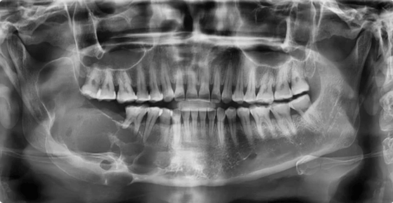
Cone Beam Computed Tomography (CBCT) is a revolutionary x-ray imaging technology, providing three-dimensional views of the patient’s dental anatomy, including teeth, bones, nerves, and surrounding tissues. Unlike traditional two-dimensional X-rays, CBCT captures high-resolution, cross-sectional images by utilizing a cone-shaped beam of ionizing radiation.
The result is a detailed and accurate representation that enables dental professionals to assess anatomical structures with precision, offering a more comprehensive understanding of the patient’s oral health. CBCT scans provide dentists and oral surgeons with valuable diagnostic information essential for accurate treatment planning and intervention when a dental tumor is suspected.

What Are Dental Tumors?
Dental tumors are abnormal growths that develop within the oral cavity, jawbones, or surrounding tissues. These tumors can originate from various structures, including the teeth, gums, bone, or soft tissues, and they may be benign (non-cancerous) or malignant (cancerous). While dental tumors are relatively uncommon compared to other oral conditions, they can present significant challenges in diagnosis and treatment due to their diverse nature and potential for affecting adjacent structures. Benign dental tumors typically grow slowly and may not cause noticeable symptoms until they reach a considerable size or impinge on nearby tissues. In contrast, malignant dental tumors can spread rapidly and pose serious health risks if not diagnosed and treated promptly. Understanding the different types, characteristics, and potential consequences of dental tumors is crucial for clinicians to effectively manage these conditions and ensure optimal patient outcomes.
What Are Different Types Of Dental Tumors?
Odontogenic tumors are a diverse group of lesions that originate from the tissues involved in tooth development. These tumors can arise from various components of the tooth-forming apparatus, including the enamel, dentin, cementum, pulp, and periodontal ligament. Understanding the different types of odontogenic tumors is essential for accurate diagnosis and appropriate management. Some common types of odontogenic tumors include:
- Ameloblastoma: Ameloblastomas are benign but locally aggressive tumors that arise from the cells responsible for forming tooth enamel. They typically occur in the mandible (lower jaw) or maxilla (upper jaw) and can cause significant bone destruction if left untreated.
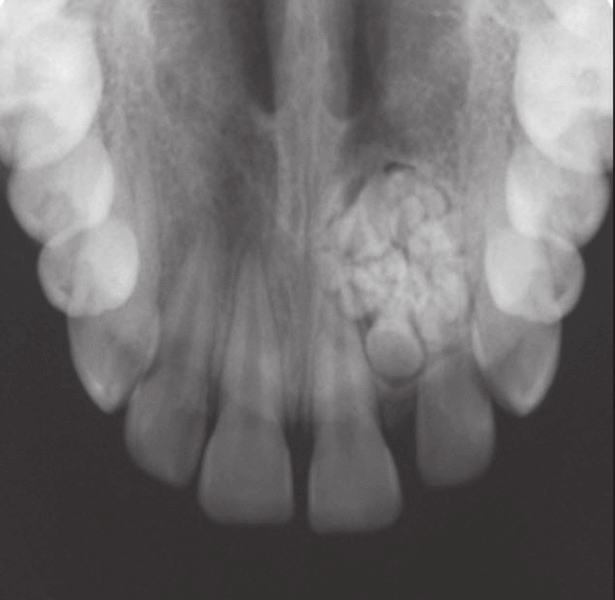
- Odontoma: Odontomas are non-aggressive, slow-growing tumors composed of dental tissues such as enamel, dentin, cementum, and pulp. They are considered developmental anomalies rather than true neoplasms and are often detected incidentally on routine dental radiographs.
- Cementoblastoma: Cementoblastomas are rare benign tumors that originate from cells responsible for forming tooth cementum. These tumors are usually attached to the roots of teeth, 9particularly molars, and are characterized by a mass of calcified tissue surrounded by a thin layer of fibrous tissue.
- Odontogenic myxoma: Odontogenic myxoma is a benign but locally invasive tumor composed of loosely arranged myxoid (mucous) connective tissue. It typically presents as a painless swelling in the jaw and may cause displacement of adjacent teeth or resorption of surrounding bone.
- Keratocystic odontogenic tumor (formerly odontogenic keratocyst): Keratocystic odontogenic tumors are cystic lesions that arise from remnants of the dental lamina. Although they are classified as cysts, their aggressive behavior and high recurrence rate warrant consideration as tumors.
- Dentigerous cyst: Dentigerous cysts are the most common type of odontogenic cysts and typically arise from the epithelial remnants associated with the formation of tooth enamel. These cysts are often associated with impacted teeth and may cause swelling, displacement of adjacent teeth, or resorption of surrounding bone.
These are just a few examples of the diverse spectrum of odontogenic tumors encountered in clinical practice. Each type of tumor presents unique clinical and radiographic features, and accurate diagnosis often requires a combination of clinical examination, imaging studies such as CBCT scans, and histopathological evaluation.
Why Do I Need A CBCT Scan For Dental Tumors?
CBCT (Cone Beam Computed Tomography) scans are indispensable tools in the evaluation of dental tumors due to their ability to provide detailed three-dimensional images of the oral and maxillofacial structures. Unlike conventional two-dimensional radiographs, CBCT scans offer superior visualization of the intricate anatomy of the jaws, teeth, and surrounding tissues, making them invaluable for detecting and characterizing dental tumors accurately.
- Comprehensive Assessment: CBCT scans allow for a comprehensive assessment of dental tumors by providing detailed information about their size, shape, location, and extent. This comprehensive evaluation is essential for determining the nature of the tumor (benign or malignant) and devising an appropriate treatment plan.
- Precise Localization: CBCT scans enable precise localization of dental tumors within the jaws, facilitating accurate surgical planning and minimizing the risk of damage to surrounding vital structures such as nerves, blood vessels, and adjacent teeth.
- Evaluation of Bone Involvement: Dental tumors often involve the surrounding bone, leading to bone destruction or remodeling. CBCT scans provide detailed images of the bone architecture, allowing clinicians to assess the extent of bone involvement and plan surgical interventions accordingly.
- Identification of Associated Complications: CBCT scans can reveal any associated complications or abnormalities caused by dental tumors, such as root resorption, cortical perforation, or sinus involvement. This information is crucial for predicting treatment outcomes and anticipating potential complications.
- Assessment of Treatment Response: Following treatment, CBCT scans can be used to evaluate the response of dental tumors to therapy, such as tumor regression, recurrence, or resolution. This ongoing monitoring helps clinicians adjust treatment plans as needed and ensures optimal patient care.
Overall, the detailed information provided by CBCT scans enhances the diagnostic accuracy, facilitates treatment planning, and improves the overall management of dental tumors. By leveraging this advanced imaging technology, clinicians can achieve better clinical outcomes and enhance patient safety and satisfaction.
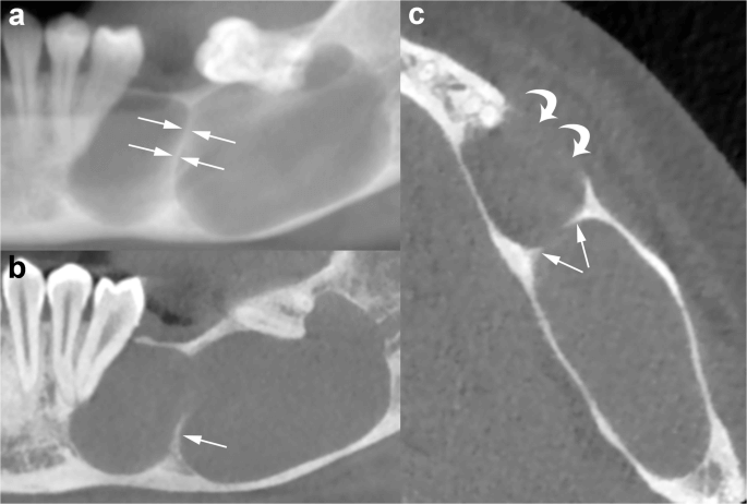
What Do Odontogenic Tumors Look Like in CBCT Scans?
Odontogenic tumors manifest in various forms within CBCT (Cone Beam Computed Tomography) scans, offering clinicians valuable insights into their morphology and characteristics. Depending on the type and nature of the tumor, CBCT images may reveal distinct features that aid in diagnosis and treatment planning:
- Radiolucent Lesions: Some odontogenic tumors appear as radiolucent (dark) lesions on CBCT scans, indicating areas of decreased density within the jawbone. These lesions may present as well-defined or irregularly shaped regions of bone resorption or cystic expansion, depending on the aggressiveness and growth pattern of the tumor.
- Radiopaque Masses: Other odontogenic tumors may manifest as radiopaque (light) masses within the jawbone, reflecting areas of increased density or calcification. These masses may exhibit varying degrees of opacity and may be associated with the deposition of mineralized tissues such as enamel, dentin, or cementum.
- Cortical Expansion: Odontogenic tumors can cause cortical expansion or thinning of the surrounding bone, which may be visualized as alterations in the contour or thickness of the cortical plates on CBCT images. Cortical expansion is often indicative of the aggressive behavior of the tumor and its potential to invade surrounding tissues.
- Root Resorption: In cases where odontogenic tumors impinge on adjacent teeth, CBCT scans may reveal evidence of root resorption, characterized by the loss of root structure or irregularities in the root surface. Root resorption is a common finding associated with aggressive tumors and may influence treatment decisions.
- Soft Tissue Involvement: Advanced odontogenic tumors may extend into the surrounding soft tissues, leading to displacement or compression of nearby structures such as the gingiva, mucosa, or muscles. CBCT scans can help delineate the extent of soft tissue involvement and guide surgical planning.
By analyzing these characteristic features in CBCT scans, clinicians can accurately identify and characterize odontogenic tumors, assess their impact on surrounding structures, and formulate optimal treatment strategies tailored to each patient’s unique condition.
How Much Does A CBCT Scan Cost For Dental Tumors?
At Oral Radiology Toronto, we provide quick and convenient dental CBCT scans for busy dentists and patients in the downtown Toronto area. Our CBCT scans are useful for assessing the teeth, their supporting structures, the mandible and maxilla up to the floor of the nose.
Our competitively priced dental CBCT scans are professionally reviewed and interpreted by a Canadian licensed Oral Radiologist.
Prices are based on the size of CBCT volume:
- Small Field CBCT (most dental cases; 5x5cm): $227
- Medium Field CBCT : $306.45
- Large Field CBCT (8x8cm): $388.50
- Panoramic X-Ray: $82
Normal CBCT report turnaround time is up to 10 business days. Expedited reporting (2 business days) is an extra $50.
Please note that we do NOT offer field of views larger than 8x8cm or imaging of structures outside the maxilla and mandible, such as the temporomandibular joints, paranasal sinuses, the cervical spine, the neck and the airway spaces (i.e. craniofacial CT scan).
How Do I Get A CBCT Scan For Dental Tumors?
- For Referring Dentists: Refer a dental patient for a CBCT Scan using our Online Referral Form.
- For Dental Patients: Schedule a CBCT scan visit at Oral Radiology Toronto using our Online Appointment Booking System.